
Fig. 101.
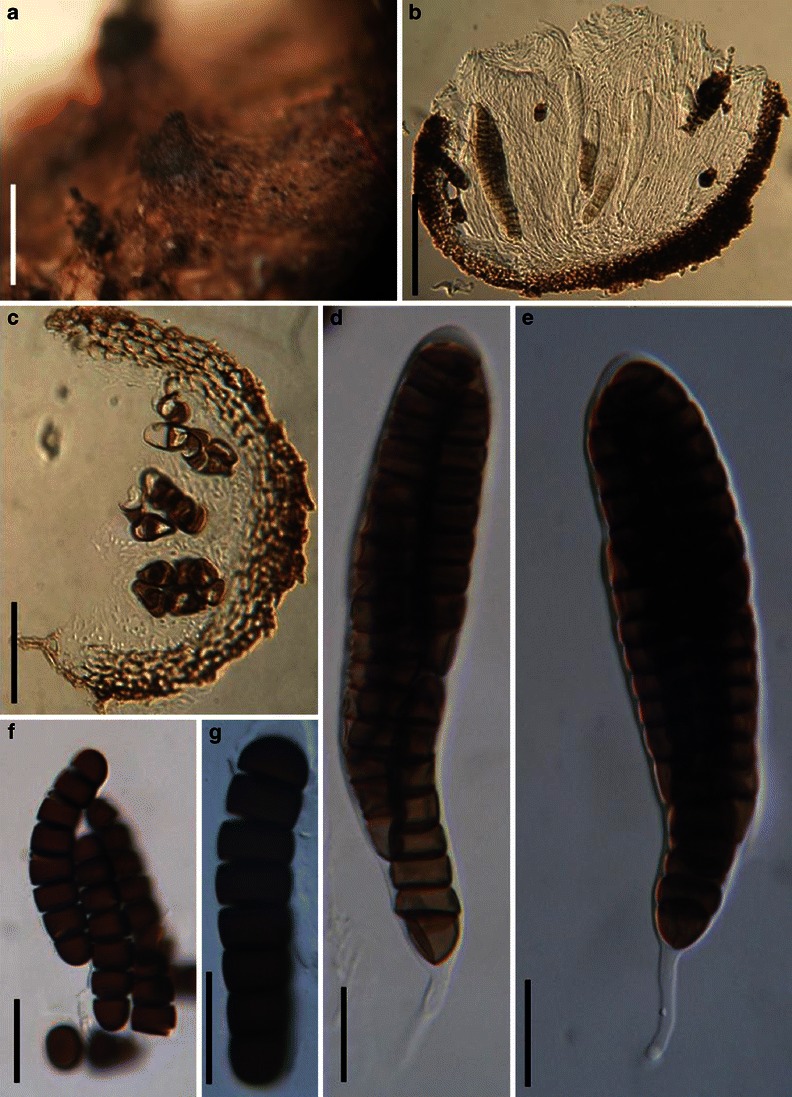
Spororminula tenerifae (from HCBS 9812, holotype). a Appearance of ascomata on the host surface. b, c Sections of the partial peridium. Note the elongate cells of textura angularis. d, e Asci with thin pedicels. f, g Ascospores, which may break into part spores. Scale bars: a = 0.5 mm, b = 100 μm, c = 50 μm, d–g = 20 μm