Fig. 12.


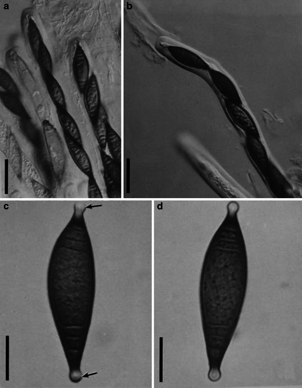
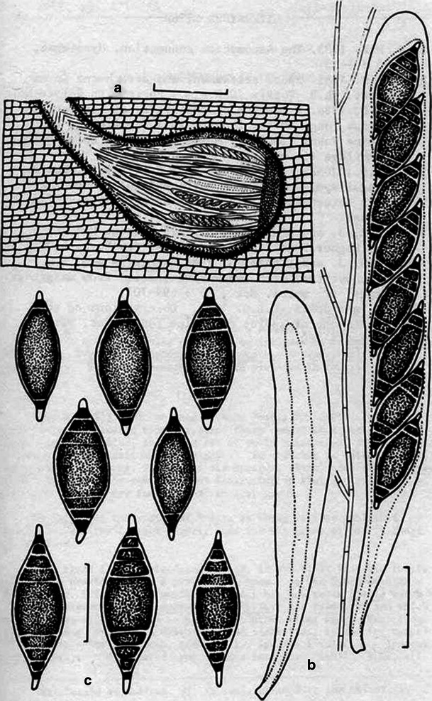
1 Biatriospora marina (from IMI 297768, holotype). a, b Cylindrical asci. Note the mucilage pseudoparaphyses in (a) and the conspicuous ocular chamber in (b). c, d Ascospores with hyaline end chambers (arrowed). Scale bars: a, b = 50 μm, c, d = 20 μm. 2 Line drawings of Biatriospora marina (based on holotype). a Section through ascocarp showing asci and pseudoparaphyses. b Asci and pseudoparaphyses. c Ascospores. Scale bars: a = 200 μm, b = 40 μm, c = 30 μm (figure with permission from Hyde and Borse 1986)
