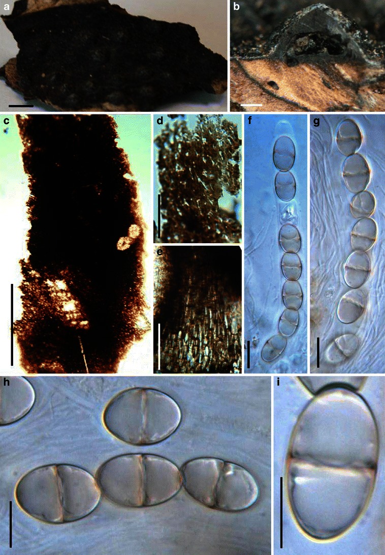
Fig. 1.

Acrocordiopsis patilii (from IMI 297769, holotype). a Ascomata on the host surface. b Section of an ascoma. c Section of lateral peridium. d Section of the apical peridium. e Section of the basal peridium. Note the paler cells of textura prismatica. f Cylindrical ascus. g Cylindrical ascus in pseudoparaphyses. h, i One-septate ascospores. Scale bars: a = 3 mm, b = 0.5 mm, c = 200 μm, d, e =50 μm, f, g = 20 μm
