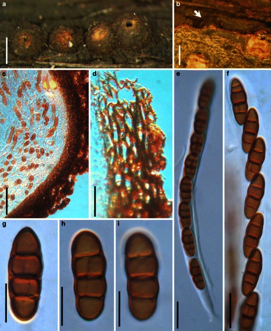
Fig. 40.

Karstenula rhodostoma (from PH 01048835, type). a Line of ascomata on host surface (after remove the decaying cover). Note the wide ostiolar opening and light colored region around the ostiole. b Immersed ascoma under the decaying cover (see arrow). c, d Section of the peridium. The peridium comprises small thick-walled cells in the outer layer. The outside comprises defuse hyphae which is probably part of the subiculum. e Ascus with a short furcate pedicel. f Partial ascus showing arrangement of ascospores. g–i Released ascospores. Note the transverse and rarely vertical septa. Scale bars: a, b = 0.5 mm, c = 50 μm, d–f = 20 μm, g–i = 10 μm
