
Fig. 54.
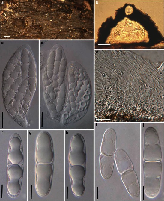
Mamillisphaeria dimorphospora (from HKU(M) 7425, paratype?). a Ascomata scattered on the host surface. Note the small papilla. b Section of an ascoma. c, d Asci (TYPE 1). e Trabeculate pseudoparaphyses in a gelatinous matrix. f–j Ascospores. Scale bars: a = 0.5 mm, b–d = 100 μm, e = 10 μm, f–j = 20 μm