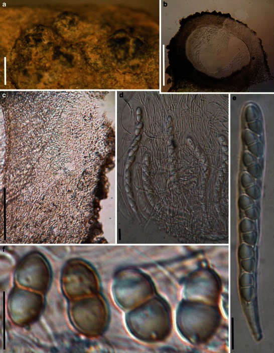
Fig. 89.

Sinodidymella verrucosa (from W 16366, type). a Ascomata on the host surface. Note the radial ridges around the pseudostiolar region. b Section of an ascoma. c Section of peridium. Note the hyaline small cells and interwoven hyphae. d Cylindrical asci in pseudoparaphyses. e Eight-spored ascus with short pedicel. f Hyaline, 1-septate ascospores which turn pale brown when mature. Scale bars: A = 1 mm, B = 100 μm, c = 50 μm, d–f = 20 μm
