Figure 1.
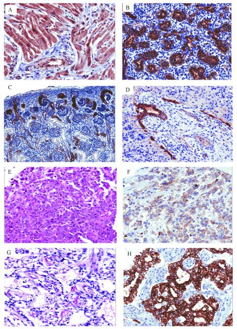
Pattern of CLDN6 expression in developing human tissues and MRT: (A) Heart (14-years-old) showing diffuse cytoplasmic staining of cardiomyocytes and smooth muscle surrounding vessels (20x). (B) Lung (22 week fetus) demonstrating strong membranous staining of alveolar lining cells (20x). (C) Kidney (19 week fetus) zone of nephrogenesis showing variable membranous staining in developing tubules (10x). (D) Liver (18 week fetus) with edge of portal triad showing strong membranous staining in developing bile ducts (20x). MRT of the liver showing (E) classic rhabdoid morphology (H&E, 20x) and (F) membranous CLDN6 staining (20x). AT/RT of the cervical spinal cord with (G) prominent vacuolar cytoplasmic changes (H&E, 20x) and (H) strong diffuse membranous CLDN6 staining (20x).

