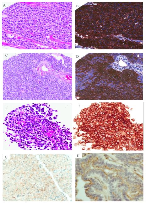
Figure 2.
Pattern of CLDN6 expression in non-MRT pediatric neoplasms: Hepatoblastoma with (A) fetal histology (H&E, 20x) and (B) strong diffuse membranous CLDN6 (20x). Wilms tumor showing (C) primitive epithelial differentiation (H&E, 20x) and (D) strong membranous CLDN6 positivity (20x). CNS germinoma with (E) classic morphology (20x, H&E) and (F) strong diffuse membranous CLDN6 staining (20x). (G) Anaplastic meningioma with focal CLDN6 staining (20X). (H) Choroid plexus papilloma showing apical CLDN6 staining simulating circumferential membranous staining in some cells cut in cross section (60x).

