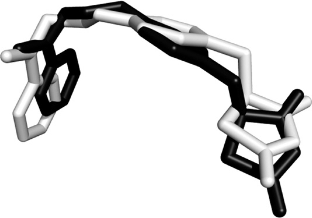
Fig. 2.
Superposition of the computed rosiglitazone binding conformation (black) docked in the PPAR-γ receptor ligand binding pocket, and the cocrystallized ligand (light gray). RMSD calculation was based on the heavy atoms, and was performed using the “rmsd” command of UCSF Chimera (http://www.cgl.ucsf.edu/chimera). PPAR-γ receptor PDB ID: 1ZGY.

