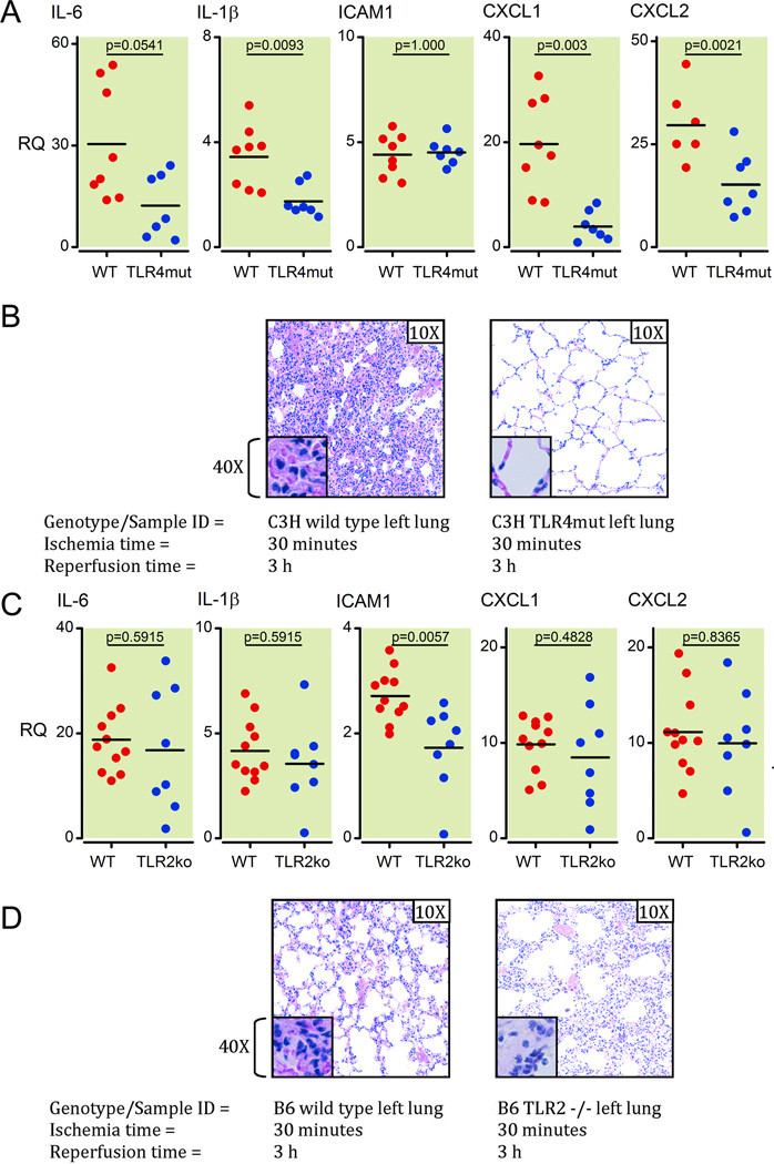
Figure 2. TLR4 presence and signaling capability is required for full pathologic response to ventilated lung I/R injury.
(A) Inflammatory cytokines and chemokine induction (mRNA) at 1 h post reperfusion measured in lung samples from C3H TLR4mut versus wild type mice using Q-PCR. All measurements were normalized to GAPDH message levels. Each point represents RNA from left lung lower segments of individual WT or C3H TLR4mut mice that underwent I/R surgery. Mice that died before completion of surgery or collection of lungs, or had inadvertent esophageal intubation and required multiple reintubation attempts were excluded from analysis (2 WT and 1 C3H TLR4mut mouse).
(B) H&E histopathology of C3H TLR4 mutant mice and C3H wild type mice left lungs at 3 h post reperfusion after I/R surgery. Each image is representative of histology images from 3 independent surgeries.
(C) Analysis of relative mRNA levels of inflammatory markers (as noted) for left lower lung segments from C57BL/6 wild type mice versus C57BL/6 TLR2 −/− mice at 1 h post reperfusion after I/R surgery. Each point represents RNA from left lung lower segments of individual WT or TLR2ko mice that underwent I/R surgery. One wild type mouse died before completion of surgery and was excluded from analysis.
(D) Histological analysis of C57/BL6 wild type versus TLR2 −/− left lungs following 30 minutes of ischemia and 3 h of reperfusion. Each image is representative of histology images from 2 independent surgeries.
TLR: toll-like receptor; mRNA: messenger RNA; Q-PCR: real-time quantitative polymerase chain reaction; GAPDH: Glyceraldehyde 3-phosphate dehydrogenase;
H&E: hematoxylin and eosin; I/R: ischemia-reperfusion; IL: interleukin; ICAM: intercellular adhesion molecule; CXCL: Chemokine (C-X-C motif) ligand; WT: wild type.

