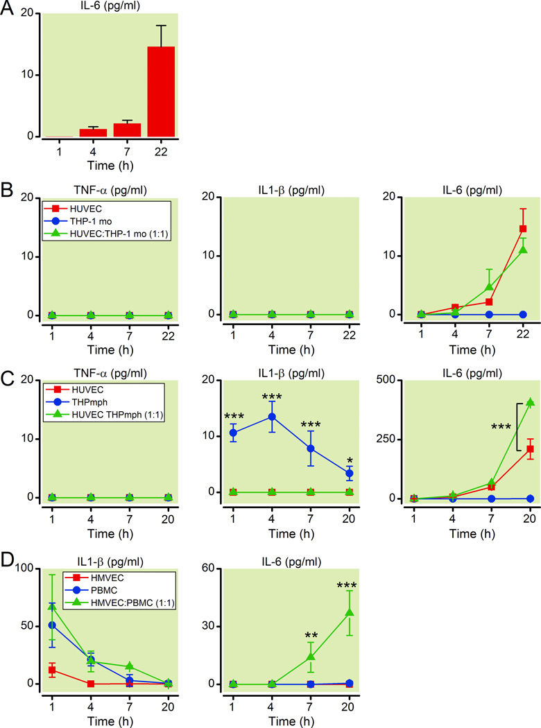
Figure 3. Generation of inflammatory cytokines from EC exposed to in vitro nutritional I/R conditions is amplified by the presence of macrophages.
(A) IL-6 protein levels were measured by ELISA at indicated time points in human umbilical vein endothelial cells (HUVEC) after they were exposed to serum-, nutrient- and glucose-free conditions for 2 h, followed by replenishment with rich complete media. Data are representative of 3 independent experiments.
(B) HUVEC were co-cultured with THP-1 human monocytes (THPmo) as noted (HUVEC or THPmo alone or HUVEC:THPmo 1:1). TNFα, IL-1β, and IL-6 protein levels were quantified from cell culture supernatants by ELISA. Data are representative of 3 independent experiments.
(C) HUVEC were co-cultured with THP-1 human differentiated macrophages (THPmph) as noted (HUVEC or THPmo alone or HUVEC:THPmo 1:1). TNFα, IL-1β, and IL-6 protein levels were assessed by ELISA from supernatants at times indicated. Data are representative of 3 independent experiments.
(D) Lung HMVEC were co-cultured with peripheral blood mononuclear cells (PBMC) as noted (HMVEC or PBMC alone or HMVEC:PBMC 1:1). IL-1β, and IL-6 protein levels were assessed by ELISA from supernatants at times indicated. Data are representative of 2 independent experiments.
EC: endothelial cells; I/R: ischemia-reperfusion; IL: interleukin; THP-1: human monocytic leukemia cell line; TNFα: tumor necrosis factor-alpha.

