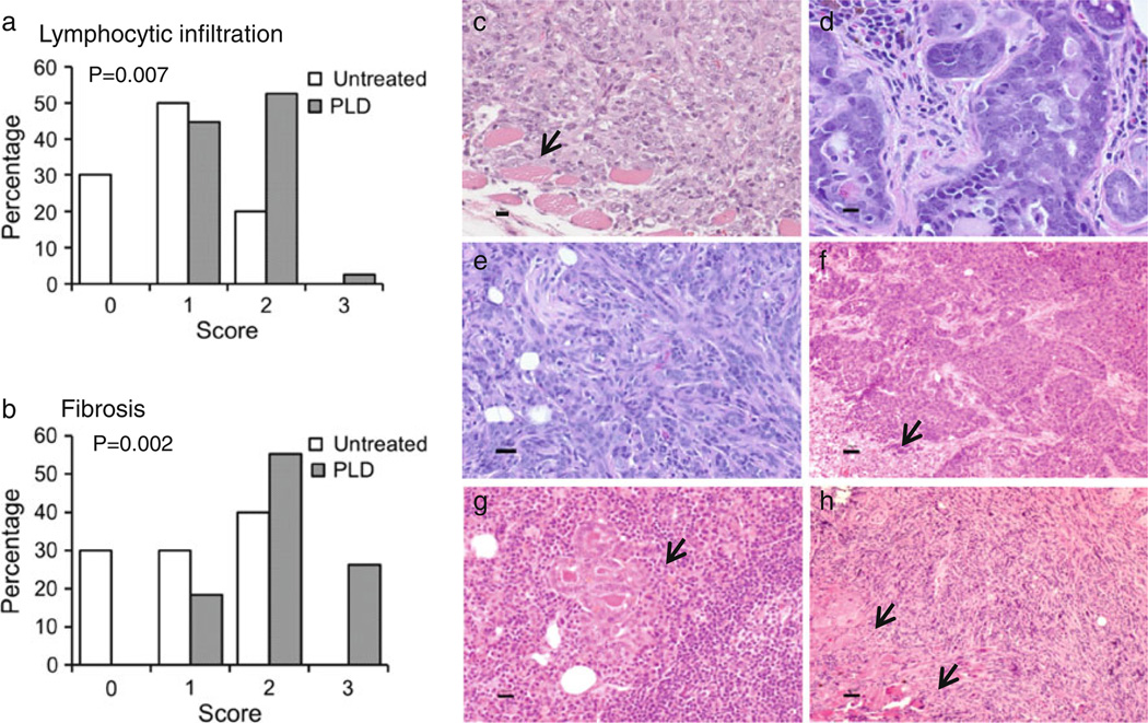
Fig. 3.
Histopathology of mammary glands and tumors in FVB/N mice. a Lymphocytic infiltration and b fibrosis were determined in each specimen. Representative histology is shown of: c invasive undifferentiated mammary carcinoma arising in mfp transplanted with epithelial cells from PLD-treated mouse, d mammary intraepithelial neoplasia, and e–h mammary tumors developing in i.duc PLD treated mice, e poorly differentiated epithelial carcinoma, f carcinoma, solid pattern with necrosis (arrow), g carcinoma invading lymph nodes (arrow) and h sarcoma-like neoplasm with EMT and local invasion of skeletal muscle (arrow). Bars 20 µm

