Figure 1.

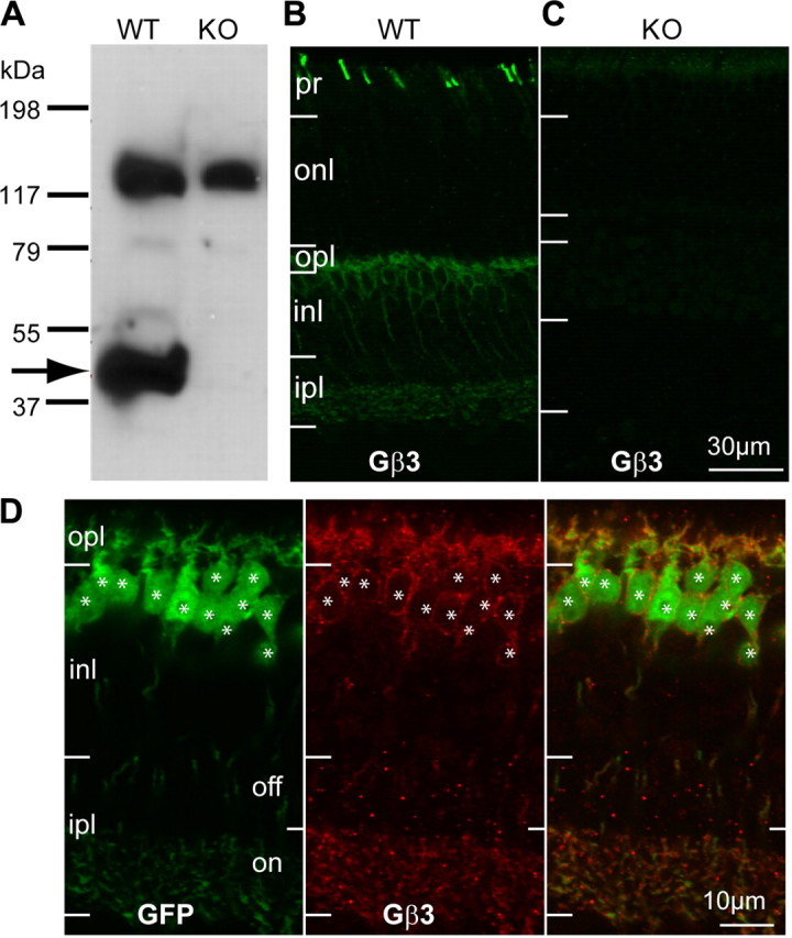
Gβ3 is expressed by cones and all types of ON bipolar cells. A, Western blots for Gβ3 in WT and Gnb3-null (KO) mice show a specific band at the expected molecular weight of 38 kDa (arrow) and a higher nonspecific band at ∼130 kDa. B, Immunostaining for Gβ3 in wild-type shows strong staining of cone outer segments (in the photoreceptor layer) and weaker staining in the outer plexiform layer and the inner nuclear layer. C, All staining is absent in the null mouse. D, Higher magnification of the stained inner retina of the Grm6-GFP mouse that expresses GFP in all and only ON bipolar cells. Strong Gβ3 staining appears in the dendrites and somas, and weak staining is present in the ON sublamina of the inner plexiform layer (ipl). Note that all GFP-expressing cells also express Gβ3 (asterisks) and vice versa. pr, Photoreceptor layer; onl, outer nuclear layer; opl, outer plexiform layer; inl, inner nuclear layer.
