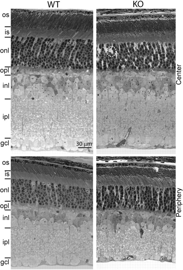
Figure 3.

Retinal layers of wild-type and Gnb3-null mice have similar appearance and thickness. Semithin (1 μm; radial view) epon radial sections of retinas (4 weeks old) stained with toluidine blue clearly show all the retinal layers. This picture is representative of three adult WTs and three adult KOs. The layers in general have a normal appearance, but in certain regions, Muller cells appear to occupy bigger spaces than normal (within the dotted circle). Top panels show the center of the retina, and bottom panels show the periphery. os, Outer segments; is, inner segments; gcl, ganglion cell layer. Other abbreviations are defined as in Figure 1.
