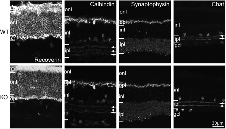
Figure 4.
Gross morphology of all cell classes is normal in the Gnb3-null retina. Immunostaining using several cell markers for WT (top) and Gnb3-null (KO; bottom) retinas. Recoverin labels all photoreceptors throughout the cell including outer and inner segments, somas, and photoreceptor terminals. All of these segments are stained in both wild-type and null retinas. Calbindin labels horizontal cells strongly and certain amacrine cells weakly. Horizontal cells and the three bands in the ipl (arrows) appear similar. Synaptophysin staining shows that the two synaptic layers are similar. Choline acetyl transferase (Chat) labels the cholinergic amacrine cells that stratify in the OFF (top band) and ON sublaminas (lower band) of the ipl; these bands are unaffected by the absence of Gβ3. Data are representative of three (synaptophysin) to eight (calbindin) images from three different animals for each genotype. Abbreviations are defined as in Figure 1.

