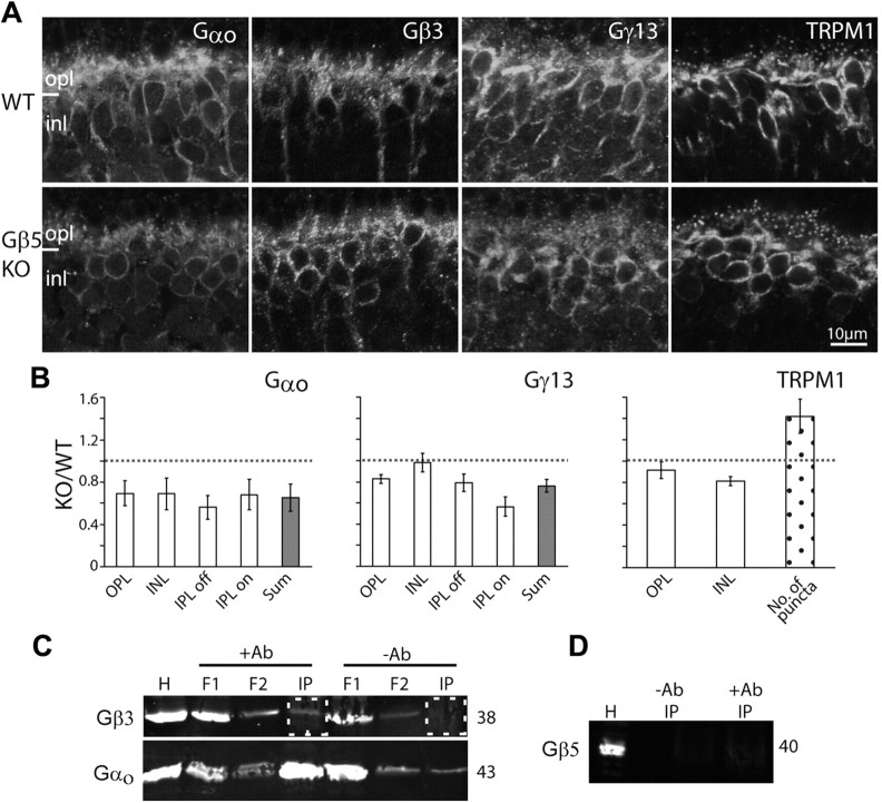
Figure 9.
Gβ3 rather than Gβ5 forms the heterotrimer Go. A, Immunostaining in wild-type (top) and Gβ5-null (bottom) retinas for Gαo, Gβ3, Gγ13, and TRPM1. Fixed retinas and sections were obtained from J. Chen. B, Relative staining intensity (as defined in Fig. 6C) for Gαo, Gγ13, and TRPM1 in several retinal layers and their sum. For TRPM1, the number of puncta was calculated as in previous figures. Gβ3 staining was not quantified because staining only worked on one set of tissues. However, results from the Chen lab showed similar staining intensity in wild-type and null tissues (personal communications). Average ratios represent results from at least three sets of experiments. C, Coimmunoprecipitation using a monoclonal antibody to pull down Gαo and its interacting proteins. The top blot was probed for Gβ3, and bottom one for Gαo. The relevant lanes, i.e., immunoprecipitated (IP) protein with and without the antibody, are boxed with dotted squares. H, Homogenate; F1, F2, flow through. D, Coimmunoprecipitation samples probed for Gβ5.

