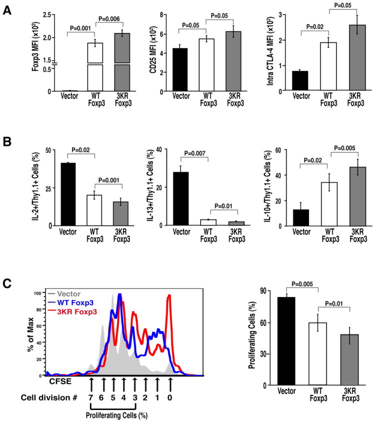
Figure 5. The suppressive function of Foxp3 is enhanced by the 3KR mutation.

(A) Flow cytometric analysis (MFI) of Foxp3, CD25, and intracellular CTLA-4 in murine CD4+ T cells transduced with retroviral vectors expressing wildtype or 3KR mutant Foxp3. (B) Flow cytometric analysis (%) of cytokine-secreting cells from transduced T cells after activation with α-CD3/CD28 antibodies. Data in (A) and (B) represent the average (mean ± SEM) of three independent experiments. (C) Suppression assays of wildtype Foxp3 or the 3KR mutant. Naive mouse CD4+ T cells were infected with retroviral vectors expressing wildtype Foxp3 or the 3KR mutant along with control retrovirus after activation by α-CD3/CD28 antibody for 24 hrs as above. Infected cells were sorted and co-cultured with CFSE-labeled responder T cells for 3 days. Proliferation of responder T cells was assessed by CFSE dilution within the responder T cell population. The left panel shows one representative FACS plot. The proliferating cells were presented as cells having divided more than 3 cycles. The right panel shows the data represented as the mean ± SEM of five independent experiments.
