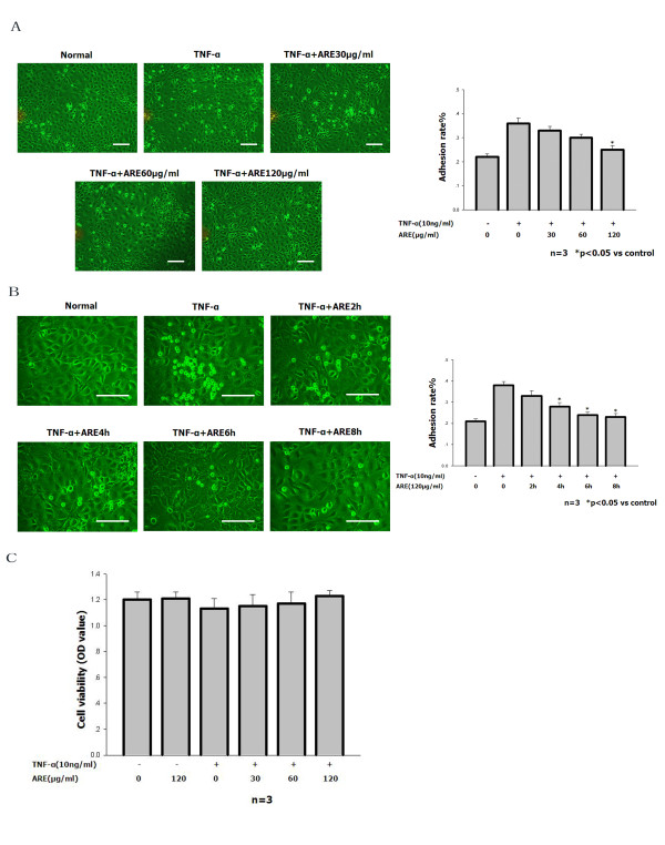
Figure 1.
ARE inhibited TNF-α-stimulated adhesion of THP-1 cells to SVEC cells. A, B. Quantitation of THP-1 cells adhering to untreated SVEC cells and to SVEC cells treated for 4 h with 10 ng/ml TNF-α in the absence or presence of ARE (30, 60 or 120 μg/ml) for up to 2, 4, 6, or 8 h. For details of the adhesion assay see Materials and methods. The number of adherent THP-1 cells was determined using a counting slide. Data are representative of at least three independent experiments and are shown as mean values ± S.D. *P < 0.05 compared with cells treated with TNF alone. Scale bars: 50 μm. C. SVEC cells were treated with 10 ng/ml TNF-α in the absence or presence of ARE (30, 60 or 120 μg/ml) for 8 h. The viability of SVEC cells was determined by MTT analysis.

