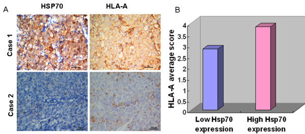
Figure 4.

Correlation between expression levels of Hsp70 and the level of HLA-A in NPC tissues.A, continuous sections of human NPC tissue were subjected to IHC staining with antibodies against Hsp70 and HLA-A. The high membranal and cytoplasmic expression levels of Hsp70 in the tumor tissue in case 1 were accompanied by an elevated level of HLA-A. Conversely, the low membranal and cytoplasmic expression levels of Hsp70 in the tumor tissue of case 2 were accompanied by a low level of HLA-A. Scale bars = 100 μm. B, in 226 NPC cases with high membranal and cytoplasmic expression levels of Hsp70, an average HLA-A score was 3.82 ( right column), an average score that was significantly higher than that (2.81) of the 281 NPCs with low expression levels of Hsp70 ( left column, P < 0.001, independent sample t-test).
