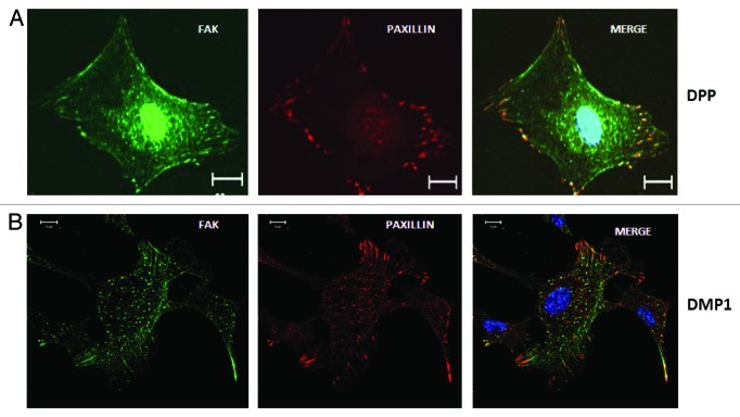
Figure 3. Localization of FAK and Paxillin in cells adherent on DPP and DMP1 coated substrate. C3H10T1/2 cells were seeded on DPP or DMP1 coated cover glass for 24 h and immunostained with anti-FAK (green) and anti-Paxillin (red) antibodies. Confocal microscopic images show successful spreading and formation of focal adhesions. Interestingly, FAK is localized in the nucleus on DPP coated substrate while it is present only at focal adhesions on DMP1 coated substrate. Paxillin is predominantly localized at the focal adhesions in cells cultured on both DPP and DMP1 substrate. Scale bars, 20 µm.

An official website of the United States government
Here's how you know
Official websites use .gov
A
.gov website belongs to an official
government organization in the United States.
Secure .gov websites use HTTPS
A lock (
) or https:// means you've safely
connected to the .gov website. Share sensitive
information only on official, secure websites.
