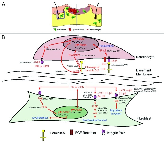
Figure 1. (A) Skin repair in wild-type mice (left) is a multistep process requiring contributions from the dermis and epidermis. Keratinocytes (pink) in the hair follicles proliferate and migrate to cover the defect. Simultaneously, fibroblasts (green) proliferate and migrate into the wound where they differentiate into myofibroblasts (red). Fibroblasts and myofibroblasts produce, secrete and organize new extracellular matrix while myofibroblasts contract the newly formed granulation tissue to draw the wound edges together. Recent reports suggest that delayed wound closure in the Postn−/− mouse (right) is due to decreased keratinocyte proliferation (leading to slowed re-epithelialization), decreased fibroblast proliferation and migration into the wound and failure of wound fibroblasts to differentiate into myofibroblasts (resulting in failed wound contraction). (B) There is considerable evidence in the literature to support a role for periostin in all of these cellular mechanisms. Predominantly, periostin has been shown to mediate its effects through integrins (such as αvβ3) and adhesive signaling.29-32 Although Nishiyama’s proposal requires intracellular interactions between periostin and BMP-1, the ability of rhPN to increase MMP2 expression, thereby facilitating laminin5γ2 cleavage, may represent an alternative mechanism for keratinocyte proliferation based on αvβ3 ligation and adhesion signaling.

An official website of the United States government
Here's how you know
Official websites use .gov
A
.gov website belongs to an official
government organization in the United States.
Secure .gov websites use HTTPS
A lock (
) or https:// means you've safely
connected to the .gov website. Share sensitive
information only on official, secure websites.
