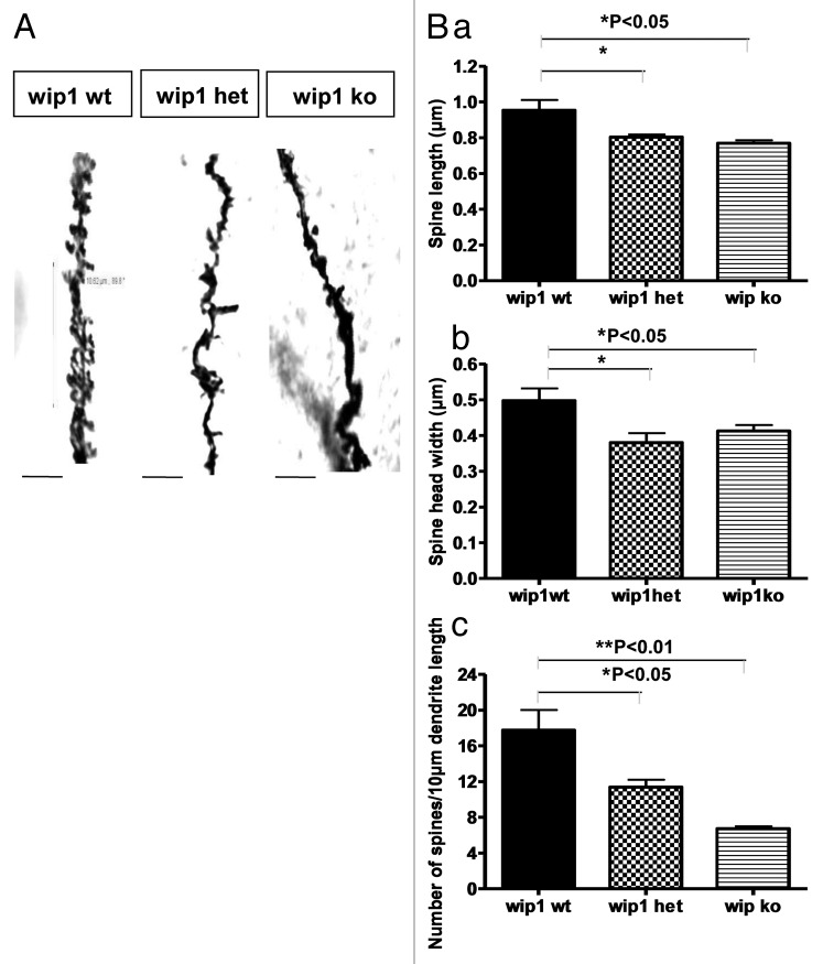
Figure 1. Morphological analysis of the dendrites in CA1 of the hippocampus in wip1 wt and wip1 ko mice. (A) Golgi staining of dendrites of adult pyramidal neurons in CA1 of the hippocampus of wip wt, wip1 het and wip1 ko mice. Representative images of dendritic fragments from wip1 wt, wip1 het and wip1 ko mice. (B) Quantification of spine length (a), spine head width (b) and spine density (c) of the Golgi stained neurons in wip1 wt (n = 9), wip1 het (n = 4) and wip1 ko mice (n = 8). *p < 0.05, **: p < 0.01. Bars = 4 ?m.

An official website of the United States government
Here's how you know
Official websites use .gov
A
.gov website belongs to an official
government organization in the United States.
Secure .gov websites use HTTPS
A lock (
) or https:// means you've safely
connected to the .gov website. Share sensitive
information only on official, secure websites.
