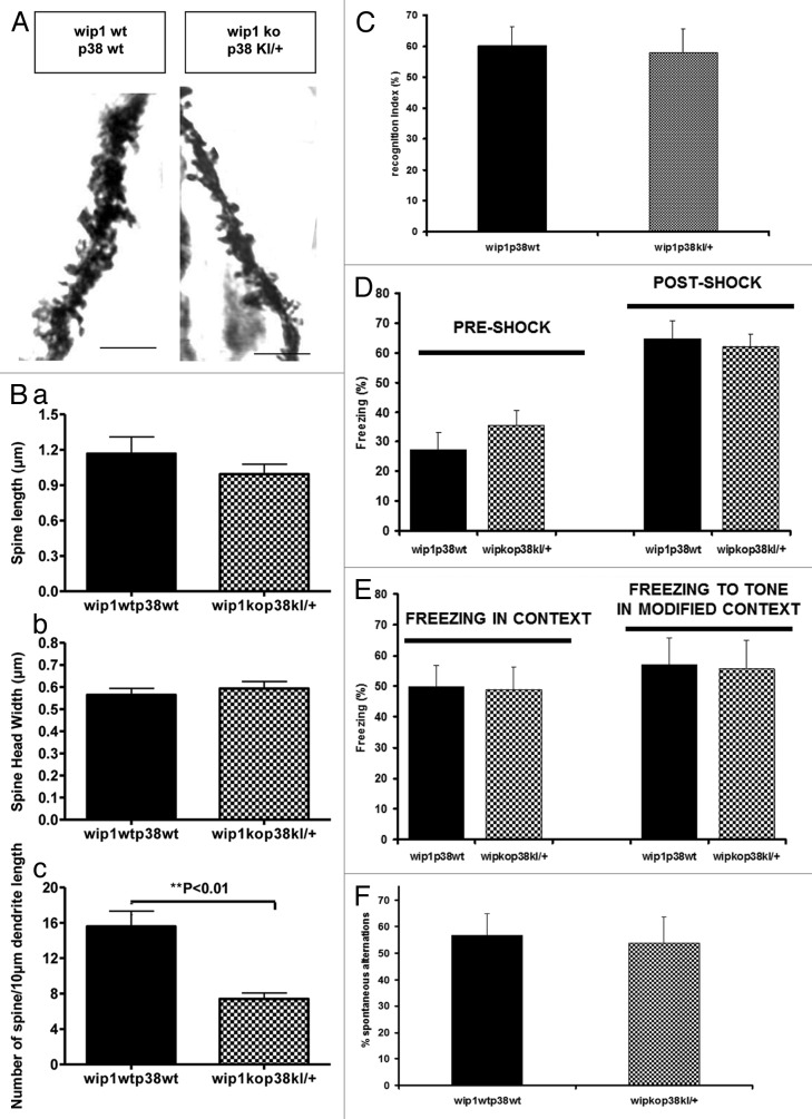
Figure 5. Wip1 deficiency-associated changes in dendritic spine morphology and behavioral tests are reversed in the wip1kop38KI/+ double mutant mice. (A) Golgi staining of dendrites of adult pyramidal neurons in CA1 hippocampus in wip wt p38wt and wip1kop38KI/+ mice. Representative pictures of dendritic fragments from wip1wtp38wt and wip1kop38KI/+ mice. (B) Quantification of spine length (a), spine head width (b), and spine density (c) of the Golgi stained neurons in wip1wtp38wt (n = 7) and wip1ko p38KI/+ mice (n = 6). Bars = 4 ?m. (C) Evaluation of memory by object recognition test in wip1wtp38wt (n = 11) and wip1kop38Kl/+ (n = 8) mice. Recognition index values are reported as mean ? SEM. (D) Freezing response of wip1wtp38wt (n = 11) and wip1kop38Kl/+ (n = 8) mice during fear conditioning trials (tone + shock conditioning). Values of freezing are expressed as mean ? SEM. (E) Freezing response of wip1wtp38wt and wip1kop38Kl/+ mice to contextual exposure and to presentation of conditioned stimulus (tone) in a modified context 24 h following fear conditioning. Values of freezing are expressed as mean ? SEM. (F) T-maze spontaneous alternation in wip1wtp38wt (n = 11) and wip1kop38Kl/+ (n = 8). Values of spontaneous alternation are expressed as mean ? SEM.

An official website of the United States government
Here's how you know
Official websites use .gov
A
.gov website belongs to an official
government organization in the United States.
Secure .gov websites use HTTPS
A lock (
) or https:// means you've safely
connected to the .gov website. Share sensitive
information only on official, secure websites.
