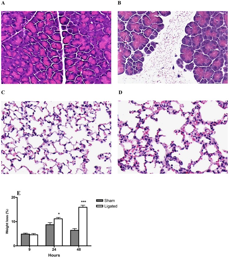
Figure 1. Morphological and physiological changes associated with acute pancreatitis and associated acute lung injury.
Representative H&E images of pancreas (A, B) and lungs (C, D) 24 hours after induction of pancreatitis through BPD ligation (A, C) are depicted. Pronounced edema and necrosis observed in the pancreas after pancreatitis induction (B) compared to sham control (A). Lung section shows thickening of alveolar septa, hemorrhage and edema after pancreatitis induction (D) compared to sham control (C). Original magnification, 20×. Mice experienced a significant weight loss 24 and 48 h after induction of pancreatitis through BPD ligation compared to sham control (E). Data expressed as mean ± SEM, n = 10 per group. *P<0.05, ***P<0.001 versus sham control, by two-tailed Student t-test.

