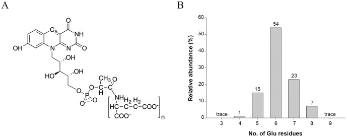
Figure 1. Molecular structure of cofactor F420.
(A) Schematic representation of cofactor F420, where n varies from 2–9 in different microorganisms. (B) Mass spectrometry analysis of cofactor F420 bound to the purified Rv0132c–Δ38 protein showing the population of species differing in the number of glutamate residues in the poly-Glu tail.

