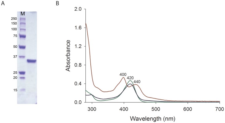
Figure 2. SDS–PAGE and UV–visible spectra of the purified Rv0132c–Δ38 protein.
(A) SDS–PAGE gel of the purified Rv0132c–Δ38 protein. (B) UV-visible spectra for purified Rv0132c (0.5 mg/mL in PBS, red), F420 extracted from M. smegmatis cells (50 µM in PBS, green) and F420 extracted from the purified Rv0132c (blue). M: molecular weight markers (kDa).

