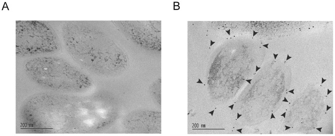
Figure 7. Immunoelectron microscopy of the M. tuberculosis H37Ra cells using anti–Rv0132c antiserum.
Electron micrographs are shown in which thin cryo–sectioned Mtb cells are (A) treated with preimmune serum, and (B) treated with anti-Rv0132c antisera at a dilution of 1/200. The gold particles (indicated by arrowheads) are present mainly on the periphery of the cells in panel B, but are absent from the control panel (A).

