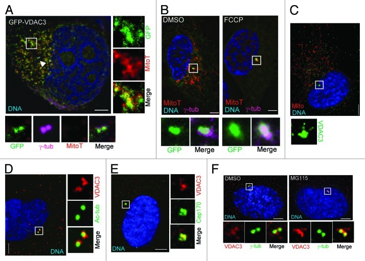
Figure 2. VDAC3 is a centrosomal protein of the mother centriole. (A) A representative HeLa cell expressing GFP-VDAC3 (green) stained with MitotrackerRed (MitoT, red), then fixed and stained for γ-tub (magenta). Arrowhead indicates centrosomes, box indicates a region containing mitochondria. (B) RPE1 cells expressing GFP-VDAC3 (green) were treated with either DMSO (solvent control) or 200 μM FCCP for 1 h and stained with MitoT (red). Shown are representative images stained for γ-tub (magenta). (C–E) Asynchronously growing RPE1 cells were stained for (C) VDAC3 (green) and mitochondria (Mito, red), (D) VDAC3 (red) and Ac-tubulin (green), and (E) VDAC3 (red) and Cep170 (green). Shown are representative images where the predominant centrosomal VDAC3 signal was at both centrosomes (C and D) or at the mother centriole (E). (F) Representative images of asynchronously growing RPE1 cells treated with either DMSO or MG115 for 4 h, stained for γ-tub (green), VDAC3 (red) and imaged under identical condition. DNA is blue and bar is 5 μm in (A–F).

An official website of the United States government
Here's how you know
Official websites use .gov
A
.gov website belongs to an official
government organization in the United States.
Secure .gov websites use HTTPS
A lock (
) or https:// means you've safely
connected to the .gov website. Share sensitive
information only on official, secure websites.
