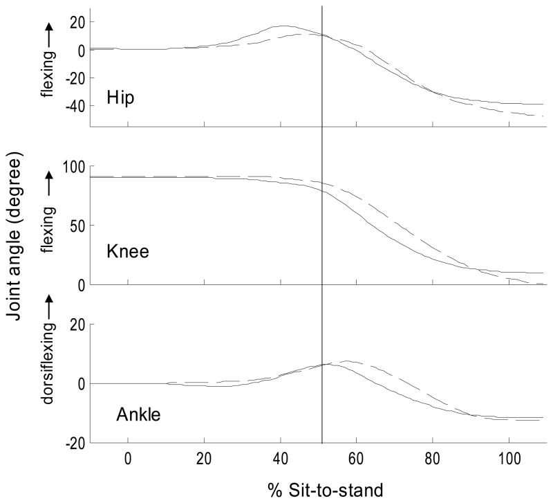
Figure 2.
Typical joint angles during the sit-to-stand. Solid line = PD-on state (ensemble-average of 5 trials for a subject with PD), Dashed line = control subject (ensemble-average of 5 trials for a control subject). The identical subjects are displayed in Figure 1. 0% = start of sit-to-stand. 100%=end of sit-to-stand. Vertical line=lift-off. For display purposes, the hip and ankle start joint angle was set to 0° and the knee at 90°. Note the hip motion was between the pelvis and thigh segment (i.e., not trunk and thigh).

