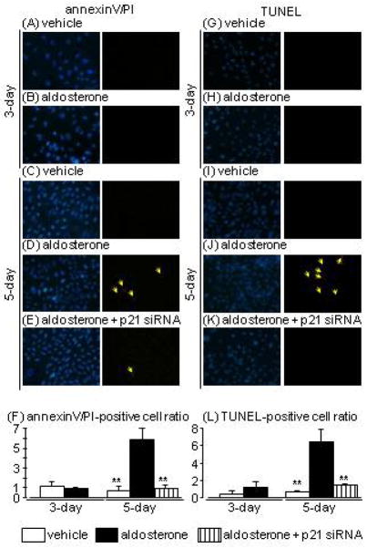
Fig. 2.
Effects of aldosterone and p21 siRNA on apoptosis evaluated by annexin-V (green)/propidium iodide (PI; red) (A–F) and terminal deoxynucleotidyl transferase dUTP nick-end labelling (TUNEL; red) staining (G–L) in human proximal tubular cells at 3 and 5 days after aldosterone treatment (arrows indicate the fluorophore-positive cells). Hoechst 33342 (blue) was used to identify nuclei in each staining (left pictures). Data are expressed as means ± S.E.M.; n = 3 or 4 per group. **P < 0.01, compared to the same-day aldosterone-treatment group.

