Figure 1.
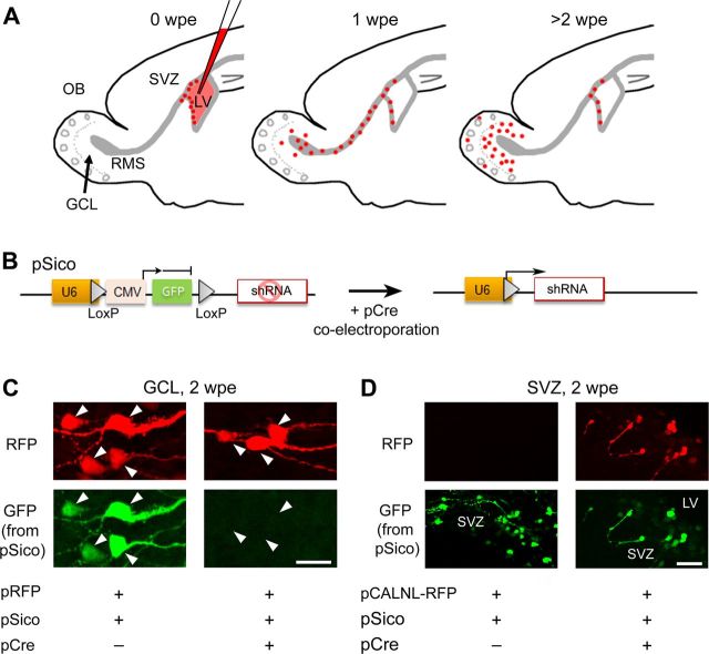
shRNA expression in the neonatal SVZ using electroporation. A, Diagrams illustrating time points and corresponding locations of SVZ-born cells following electroporation at P0–P1. B, Diagram of pSico before and after Cre-mediated recombination and gfp excision allowing shRNA expression. C, Confocal images of granule neurons expressing pRFP and pSico with or without pCre coelectroporation. Loss of GFP expression with Cre suggests functional recombination of the plasmid. D, SVZ cells expressing pCALNL and pSico with or without pCre coelectroporation. Note that the GFP from pSico persists in the SVZ despite functional Cre recombination as indicated by the presence of RFP from pCALNL. Scale bars: C, 25 μm; D, 50 μm.

