Fig. 2.

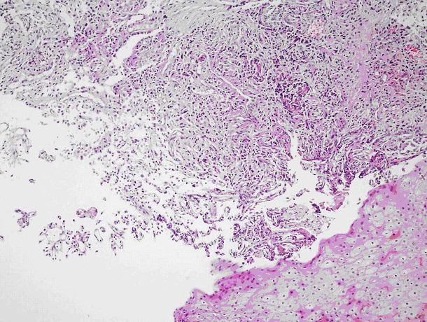
A tumour manifesting an alveolar pattern separated by long, thin vascular structures formed by uniform, round cell nuclei with granular cytoplasm under regular, multi-layered squamous epithelium (hematoxylin and eosin stain ×240).

A tumour manifesting an alveolar pattern separated by long, thin vascular structures formed by uniform, round cell nuclei with granular cytoplasm under regular, multi-layered squamous epithelium (hematoxylin and eosin stain ×240).