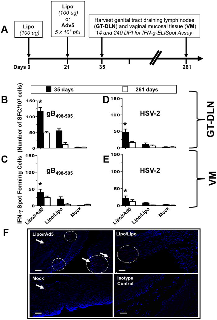
Figure 1. Potent and long-lasting CD8+ T cell responses detected in both GT-DLN and VM following IVAG immunization with Lipo/rAdv5 prime/boost vaccine.

(A) Time course for immunization and CD8+ T cell response analysis. Four groups of B6 mice (n =10) were immunized IVAG with: 100 μg of lipopeptide in saline on days 0, and 21 (Lipo/Lipo), with lipopeptide on day 0 and then with 5 × 10⁷ pfu of rAd5 in saline on days 21 (Lipo/rAdv5 prime/boost mucosal vaccine or Lipo/rAd5), or with the irrelevant OVA257-264 lipopeptide and an empty Ad5 vector in saline on days 0 and 21 (mock-immunized or Mock). (B–E) On day 35 and 261 (i.e. fourteen and 240 days post-immunization (DPI)), the iliac and inguinal lymph nodes draining the genital tract (GT-DLN) (B and D) as well as the vaginal mucosa (VM) (C and E) were harvested. GT-DLN and VM cell suspensions were assayed for gB498-505- (B and C) and HSV-2- (D and E) specific IFN-γ-producing CD8+ T cell responses using ELISpot assay, as described in Material & Methods. Values represent the mean of IFN-γ spot forming cells detected in an average of 5 mice. (*) Indicates the P values were ≤ 0.05 when HSV-2 or gB498-505-specific IFN-γ-secreting CD8+ T cell responses from the Lipo/rAdv5 vaccine group of mice was compared to the homologous Lipo/Lipo group or to the mock-immunized group (one-way ANOVA). (F) Vaginal mucosal tissue was collected 14 days after the second immunization from the progesterone-treated mice that were immunized with Lipo/rAdv5 (upper left), Lipo/Lipo (upper right) or mock-immunized (lower left) and CD8+ T cells were detected by immunofluorescence microscopy, as described in Material & Methods. Sections were stained with fluorescein isothiocyanate-conjugated anti-CD8 antibody, and the nuclei were visualized by staining with DAPI (blue). Arrowheads and circles indicate CD8+ T cells accumulating at the vaginal epithelium and stroma. Staining with an isotype control IgG is shown in the lower right picture. Scale bars = 50 υm. Results are representative of two independent experiments.
