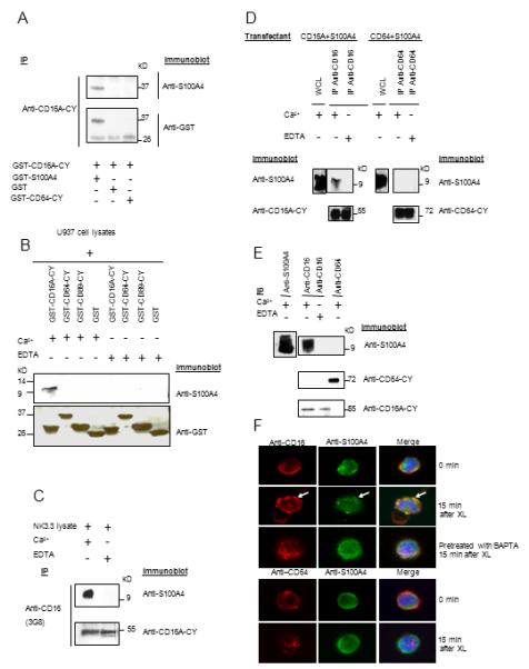
FIGURE 6.

The interaction of CD16A-CY with S100A4 is direct, specific and calcium-dependent. (A) GST-CD16A-CY in binding buffer containing 1 mM CaCl2 or 10 mM EDTA, was incubated with GST-S100A4, GST-CD64-CY, or GST. GST-CD16A-CY was precipitated with polyclonal anti-CD16A-CY and sequentially blotted with polyclonal anti-S100A4 and anti-GST (n=3). (B) U937 cell lysates were incubated with GST-CD16A-CY, GST-CD64-CY, GST-CD89-CY, or GST on glutahione-Sepharose 4B beads in the presence of 1mM Ca2+ or 10 mM EDTA overnight at 4°C. The beads were washed and the proteins were resolved and immunoblotted sequentially with anti-S100A4 and anti-GST (n=3). (C) Interaction between CD16A and S100A4 in NK3.3 cell line in the presence of 1mM Ca2+ or 10mM EDTA. Cell lysates were immunoprecipitated with 3G8 and analyzed by sequential immunoblotting with anti-S100A4 and anti-CD16-CY (n=3). (D) 293 cells transiently expressing S100A4 and CD16A or CD64 were lysed with NP40 lysis buffer with or without Ca2+ 48 hr after transfection. CD16A and CD64 immunoprecipitates were sequentially blotted with anti-S100A4, anti-CD16-CY, and anti-CD64. S100A4 expression in the cells was examined by immunoblotting with anti-S100A4 in the cell lysate (upper panels) (n=3). (E) Freshly prepared mononuclear cells were lysed with NP40 lysis buffer with 1mM Ca2+ or 10 mM EDTA. CD16A or CD64 immunoprecipitates were blotted with anti-S100A4, anti-CD16-CY, and anti-CD64-CY sequentially. S100A4 expression in the cells was examined by immunoblotting with anti-S100A4 in the cell lysate (upper left panel) (n=3). (F) Mononuclear cells from donors were incubated with Alexa Fluor 594 conjugated 3G8 F(ab’)2 (10 ug/ml) for 40 min at 4°C to label surface CD16 (upper 3 panels). Cells were then washed with PBS and cross linked at 37°C for 15 min. Cells were fixed with 4% paraformaldehyde in PBS for 20 min at room temperature and permeabilized with 0.2 % Triton in PBS for 2 min at room temperature. Cells were incubated with Alexa Fluor 488 conjugated anti-S100A4 for 1 hr at room temperature. Alternatively, the cells were pre-treated with 10 mM BAPTA-AM for 30 min at 37°C before cross-linking and staining. Mononuclear cells were incubated with Alexa Fluor 594 conjugated anti-CD64 for 40 min at 4°C to label surface CD64, cross-linked and analyzed as above. Data are representative of 6 experiments.
