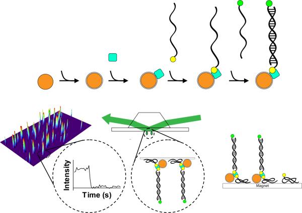
Figure 1.
Schematic for the assembly, directed immobilization, and single-molecule detection of captured DNA targets by magnetic nanoparticles. 1) The Fe3O4 nanoparticles are prepared by the coprecipitation method. 2) The nanoparticles are treated with APTES to activate the surface. 3) Streptavidin molecules (filled blue square) are conjugated to the surface via the EDAC-mediated coupling reaction. 4) The DNA recognition site is attached through a biotin-streptavidin linkage (biotin is indicated by a filled yellow circle). 5) The dye-labeled target DNA is captured by hybridization. The Cy3 dye is shown as a filled green circle. 6) The nanoparticle assembly is added to the flow chamber, and a magnet is applied to attract the nanoparticles to the BSA-biotin-treated surface, thus reducing the search volume. 7) The immobilized nanoparticles are detected by single-molecule TIRFM. 8) Within a single field of view, the fluorescence signals from individual molecules are acquired. The inset shows the time trace for the indicated molecule. The stepwise decrease in fluorescence corresponds to dye photobleaching.

