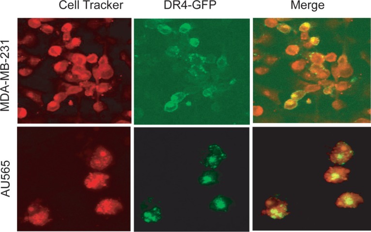
Figure 2. Confocal microscopy images showing the subcellular localization of ectopically expressed GFP-DR4 protein.
Cells were transiently transfected with GFP-DR4 expression plasmid (green), and counterstained with CellTracker Red (red). Images shown are representative of three independent experiments.

