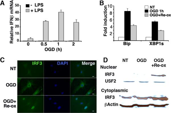
Figure 1.

Oxygen glucose deprivation (OGD) synergistically augments LPS-induced IFN-β and causes IRF3 nuclear translocation. A) MEFs were subjected to 0, 0.5, 1, or 2 h OGD, followed by media with, or without 10ng/mL LPS for 3h in standard culture conditions. IFN-β expression was determined by quantitative PCR (qPCR) with normalization to 18S rRNA (Relative mRNA). Bars represent combined results from 3 independent experiments with error bars showing SEM. p<0.0005 comparing OGD pretreated conditions and no OGD. Comparable results have been obtained in 5 independent experiments. B) MEFs were subjected to 1h OGD followed by standard culture conditions as above. Expression of BiP and spliced XBP1 was detected by qPCR and normalized to untreated controls for fold induction (NT=1). Results are representative of 2 individual experiments and error bars denote S.D. P<0.0004 comparing OGD treated and NT cells. C) MEFs were subjected to OGD for 1h followed by 2h re-oxygenation (Re-ox) in standard culture as indicated. Fixed cells were incubated with anti-IRF3 followed by anti-rabbit IgG Alexa Fluor 488, and visualized by immunofluorescence microscopy. Results are representative of 3 independent experiments. D) Nuclear and cytoplasmic lysates were resolved by SDS PAGE and immunoblotted with anti-IRF3, β-actin, or the nuclear protein anti-USF2. Results are representative of 2 separate experiments.
