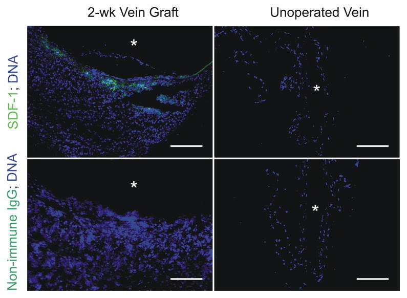
Fig 2.
SDF-1 is expressed in arterializing vein grafts. Carotid interposition vein grafts in WT mice were created with syngeneic inferior venae cavae and harvested 2 wk post-op, before the completion of arterialization. IVCs from WT mice of equivalent age were also harvested, for comparison. Serial frozen sections were stained with SDF-1-specific or non-immune rabbit IgG, anti-rabbit IgG/Alexa-488 (green), and Hoechst 33342 to visualize DNA (blue). Shown are sections from single specimens, representative of 4 independent vein graft and IVC specimens. Scale bar = 100 μm (original mag nification ×220). Equivalent results were obtained with WT/WT, WT/Cxcr4−/+, and Cxcr4−/+/Cxcr4−/+ vein grafts (see Methods).

