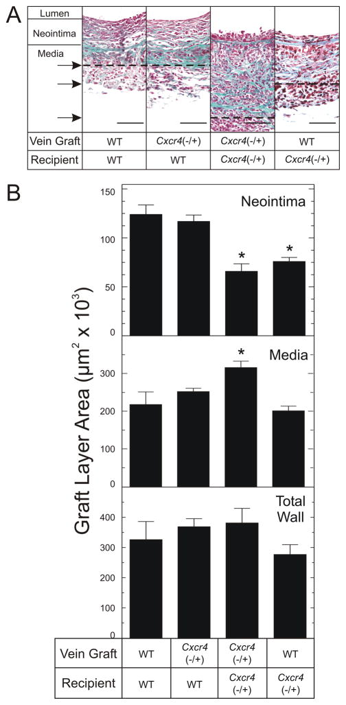
Fig 3.
CXCR4 activity in vein graft-extrinsic cells contributes to vein graft neointimal hyperplasia. Carotid interposition vein grafts from the indicated mouse donors were placed into the indicated congenic recipient mice and harvested 6 wks later, after perfusion fixation. Vein graft sections were stained with a modified connective tissue stain. A, Photomicrographs of single specimens, representative of ≥ 5 obtained of each type. Specimens are aligned at the neointimal/medial border; the medial/adventitial borders are indicated by the arrows and corresponding dashed lines. Scale bar = 50 μm (original magnification ×440). B, Neointimal and medial areas were measured for each specimen cross section by an observer blinded to specimen identity, using computerized planimetry. “Total Wall” refers to the sum of neointimal plus medial areas. Shown are means ± S.E. from ≥ 5 independent vein graft specimens from each group. Compared with WT/WT control vein grafts: *, P < .05.

