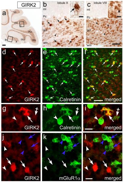
Figure 12. The brushes of CR+ UBCs are intensely labeled by GIRK2 antibody.
ac, Images from deparaffinized, DAB immunoreacted parasagittal sections of wild-type mouse cerebellum. At low magnification GIRK2 immunoperoxidase labeling is evenly localized to the granular layer across the cerebellar lobules. In addition, the granular layer of folium IXc and lobule X contains GIRK2-positive UBCs, whose brushes are much more intensely labeled than the parent cell bodies. (arrows in panel b and inset b). Granule cells are much less intensely anti-GIRK2 immunoreative in lobule X and folium IXc (compare panel b to panel c) than in other vermal lobules. Panels b and c show boxed areas from panel a. Molecular layer, ml; Purkinje cell layer, Pc; white matter, wm. d-l, Confocal immunofluorescence images from cerebellar cryosections of wild type (d-i) and Tg(Grp-EGFP) mice (j-l). Intense GIRK2 immunoreactivity is present in the brushes (white arrows) of CR+ UBCs (white arrowheads indicate UBCs cell bodies), By contrast, mGluR1 + UBCs are at background level (blue arrowheads and arrows in panels j-l). Crossed-arrows in panels g-i, point to a CR+/GIRK2-negative mossy fiber terminal. Scale bars: a, 200 μm; b-f, 50 μm; g-l and insets in b, c, 10 μm.

