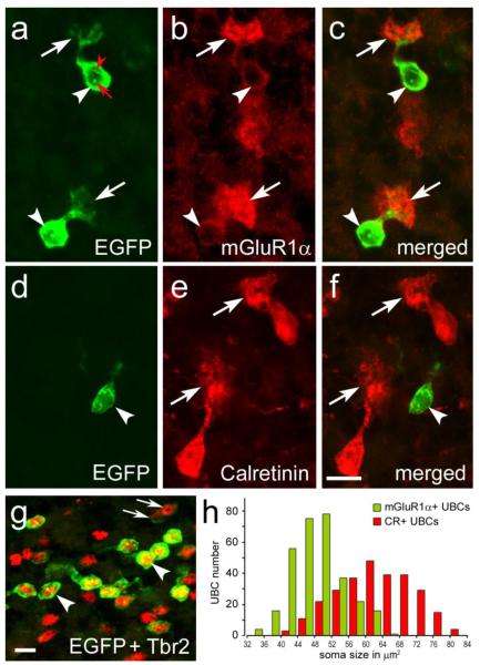
Figure 3. Immunofluorescence of chemically distinct UBC subclasses in the cerebellum of Tg(Grp-EGFP) BAC-transgenic mice.
a-g, Confocal images from two-color immunolabeled cryosections. EGFP-positive UBCs are mGluR1α+ (a-c), but CR-negative (d-f). In panels a-f arrowheads indicate cell bodies and arrows point to brushes; the nucleoli of EGFP-positive UBCs are conspicuously labeled (red arrow) and a nuclear infolding is labeled by red arrowhead. EGFP is intensely expressed in many UBCs labeled by Trb2 (arrowhead); faintly EGFP-positive UBCs are indicated by arrows (g). h, Bar graph illustrates cell subclass differences in the soma size of mGluR1 + and CR+ UBCs. Scale bars: a-g, 10 μm.

