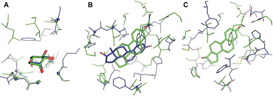
Figure 9.
The structure alignment performance of G-LoSA against evolutionary non-related proteins. (A) BS-structures of Artocarpus Integer artocarpin (PDB:1VBP) and Pterocarpus Angolensis lectin (PDB:1Q8Q), aligned by G-LoSA. (B) BS-structures of human estrogen receptor α ligand-binding domain (PDB:1A52) and human 17-β-hydroxysteroid-dehydrogenase type 1 (PDB:1FDT), aligned by G-LoSA. (C) The same structures to Figure B, but the structures are superposed using the ligand coordinates. In Figure A and B, the aligned Cα-atoms (inter-Cα distance ≤ 2.5 Å) are represented as spheres.

