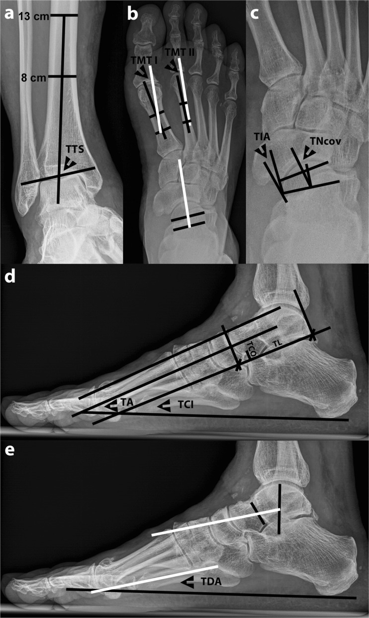
Fig. 1.
Weight-bearing radiography of a patient with varus ankle osteoarthritis showing the radiographic measurements to determine the three-dimensional position of the talus. The following measurements were performed: a Frontal tibiotalar surface angle (TTS): the mid-longitudinal tibial axis was formed by a line bisecting the tibia at 8 and 13 cm above the tibial plafond. b Horizontal talometatarsal I and II angles (TMT I and II): all white lines run parallel and represent the talar axis. c Talonavicular coverage angle (TNcov) and talonavicular incongruency angle (TIA). d Talar angle (TA), talocalcaneal inclination angle (TCI), talocalcaneal overlap (TCO), and talar length (TL). e Talar declination angle (TDA): both white lines run parallel and represent the talar axis. The tarsal index in d was determined by using TCI, TCO, and TL

