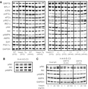
FIG. 4.
Western blot analyses of liver and cultured cells. A: Western blot analyses of the livers from WT, vaspin Tg, vaspin+/+, vaspin+/−, and vaspin−/− mice fed standard (STD) and HFHS chow. Upregulation of pAkt and pAMPK was noted in vaspin Tg mice, and pAkt and pAMPK were downregulated in vaspin−/− HFHS mice. ATF6, pelF2α, and pIRE1α were upregulated in vaspin−/− HFHS mice. B: In H-4-II-E-C3 cells, pAkt and pAMPK were enhanced by treatment of rhVaspin. C: Non-target shRNA control lentivirus transduction particles upregulated basal activity of pAkt; however, treatment of shRNA lentivirus for MTJ-1 and GRP78 suppressed pAkt in H-4-II-E-C3 cells. Impact on pAMPK was minimal by the treatment of shRNA lentivirus for MTJ-1 and GRP78. GAPDH, glyceraldehyde-3-phosphate dehydrogenase.

