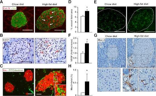
FIG. 1.
Hypervascularization and macrophage infiltration in pancreatic islets from insulin-resistant HFD-fed mice. A: Islet vessels were revealed by dual collagen IV (red) and insulin (green) immunostaining. Arrows point to thickened basement membrane. B: Endothelial cells were immunostained by CD31 antibody. HFD-fed mice showed more endothelial cells per islet (arrows). C: Pancreatic sections from HFD- and chow-fed FITC-dextran–infused mice showing vessels (green) and insulin (red). Capillaries with a large lumen are shown in the inset. D: HFD animals (black bar) showed increased FITC-dextran area/islet area compared with chow-fed mice (white bar) (n = 3/group). *P < 0.05 HFD vs. chow diet. E: VEGF immunostaining of pancreatic sections from HFD- and chow-fed mice. F: VEGF content in protein extracts of isolated islets from individual animals chow-fed mice (white bar) and HFD mice (black bar) was determined by ELISA (n = 4/group). HFD mice displayed a marked increase in Mac-2–positive macrophage infiltration in islets as shown after immunohistochemical analysis (G) and morphometrical analysis (H) (chow-fed mice [white bar] and HFD mice [black bar]). Infiltrating macrophages at the periphery and inside the islet are shown in the inset (arrows) (n = 3/group). Scale bars, 100 µm. *P < 0.05 HFD vs. chow. (A high-quality digital representation of this figure is available in the online issue.)

