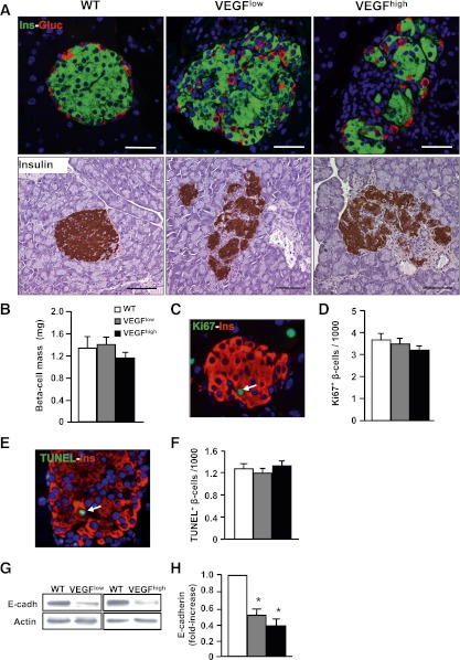
FIG. 3.
Two-month-old VEGFlow and VEGFhigh mice showed disorganization of islet architecture, but normal β-cell mass. A: Top panel: immunohistochemical analysis of insulin (green) and glucagon (red). In transgenic mice, islets appeared disorganized with α-cells in the core. Bottom panel: insulin immunostaining used to visualize islet architecture. B: β-Cell mass was measured in 2-month-old mice (n = 4–7/group): wild-type (WT) mice (white bar), transgenic VEGFlow mice (gray bar), and transgenic VEGFhigh mice (black bar). C: Ki67 (green) and insulin (red) immunostaining were used to label proliferating β-cells (arrow). D: Percentage of Ki67-positive (replicative) β-cells. E: TUNEL (green) and insulin (red) immunostaining showed apoptotic nuclei (arrow). F: Quantification of TUNEL-positive (apoptotic) β-cells. G and H: Western blot analysis of E-cadherin using islet homogenates from 2-month-old mice. G: A representative immunoblot is shown. H: Densitometric analysis of three different immunoblots: WT mice (white bar), transgenic VEGFlow mice (gray bar), and transgenic VEGFhigh mice (black bar). Scale bars, 100 µm. *P < 0.05 transgenic vs. WT. (A high-quality digital representation of this figure is available in the online issue.)

