FIG. 7.
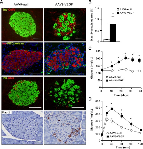
AAV-mediated VEGF overexpression in β-cells increased islet vascularization and inflammation. Two-month-old wild-type (WT) mice were injected with VEGF-expressing (AAV9-VEGF) or nonexpressing (null) AAV9 vectors (1012 vector genomes/mouse). A: Ten days after AAV injection vasculature structure was revealed by immunostaining for collagen IV (red) and insulin (green) (top panel). VEGF-treated islets showed increased basement membrane compared with AAV-null. FITC-dextran (green) together with insulin (red) immunostaining was used to label functional blood vessels (top middle panel). Insulin (green) and glucagon (red) expression showed islet disorganization (bottom middle panel). Macrophage infiltration in AAV-VEGF–treated animals was determined by Mac-2 immunostaining 10 days after AAV injection (bottom panel). Scale bars, 100 µm. B: AAV-VEGF–injected animals showed increased Mac-2–positive area/islet area when compared with AAV-null–treated mice as early as 10 days after injection: AAV-null–treated mice (white bar) and AAV-VEGF–treated mice (black bar) (n = 4 mice/group). C: Fed blood glucose levels were determined before AAV injection (day 0) and at several time points thereafter: AAV-null-treated mice (white circle) and AAV-VEGF–treated mice (black square) (n = 10 mice/group). D: Glucose tolerance was measured 20 days after AAV administration (2 g/kg body weight) (n = 10 animals/group). *P < 0.05 VEGF vs. null. (A high-quality digital representation of this figure is available in the online issue.)

