Fig. 1.
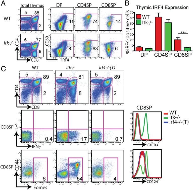
IRF4 is up-regulated during thymic selection but is not required for normal T-cell development. (A) Thymocytes from WT or Itk−/− mice were isolated and surface-stained for CD4, CD8, and CD69, and then for intracellular IRF4 protein. Thymocyte subsets (DP, CD4SP, and CD8SP) were gated on, and IRF4 vs. CD69 is displayed. Numbers on dot-plots represent the percentage of cells expressing IRF4, based on comparison with an isotype control. Data are representative of six or more independent experiments. (B) Compilation of the percentage of IRF4-expressing cells for each thymocyte subset (n = 15; ***P < 0.001 based on the Mann–Whitney test). (C) Thymocytes from WT, Itk−/−, and Irf4−/−(T) mice were stained with antibodies to CD4, CD8, CD44, intracellular Eomes, CXCR3, and CD124; stimulated for 4 h with phorbol 12-myristate 13-acetone (PMA) and ionomycin; and stained for intracellular IL-4 and IFN-γ. CD4 vs. CD8 profiles are shown (Top) along with gated CD8SP cells (Middle and Bottom).

