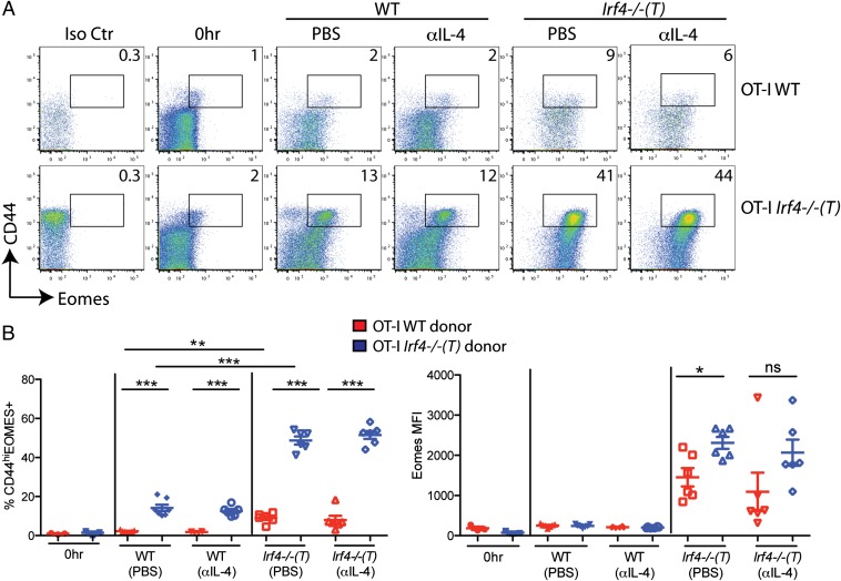
Fig. 4.
High expression of Eomes by IRF4-deficient CD8+ T cells is dependent on both cell-intrinsic and cell-extrinsic factors in Irf4−/−(T) mice. Splenocytes from OT-I WT and OT-I Irf4−/−(T) mice were mixed 1:1 and adoptively transferred into WT or Irf4−/−(T) hosts and then injected i.p. with 1 mg of αIL-4 neutralizing antibody or PBS 2–4 h before adoptive transfer. CD8+ T cells were analyzed 44 h later with a viability stain, along with antibodies to CD8, Vα2, Vβ5, CD45.1, CD45.2, CD44, CD62L, and intracellular Eomes or an isotype control. (A) Dot-plots show Eomes vs. CD44 staining on gated live CD8+, Vα2+, and Vβ5+ cells distinguished by CD45.1 (WT) vs. CD45.2 [Irf4−/− (T)+] staining. Numbers indicate the percentages of CD44hiEomes+ cells. Iso Ctr, antibody control for Eomes staining; 0 h, cells analyzed directly ex vivo before adoptive transfer. (B) Compilations of data show percentages of CD44hiEomes+ cells (Left) and raw MFI of Eomes staining (Right) on live CD8+, Vα2+, and Vβ5+ cells distinguished by CD45.1 (WT) vs. CD45.2 [Irf4−/−(T)+] staining from three independent experiments with two to three host mice per group per experiment. Statistical significance was determined by a t test (*P = 0.01; **P = 0.001; ***P < 0.0001). ns, not significant.

