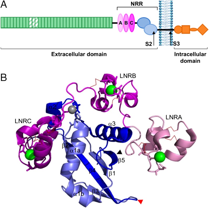
Fig. 1.
The Notch receptor and the NRR of hN2. (A) The domain structure of the Notch receptor contains an extracellular region, consisting of the NRR and the EGF-like repeats (green; repeats required for ligand interactions are striped). The NRR comprises three LNR modules (light pink to dark pink) and the furin-cleaved HD domain (HD-N, dark blue; HD-C, light blue). Approximate positions of S2 and S3 cleavage sites, which release the intracellular domain, are shown. (B) X-ray crystal structure of hN2-NRR in its autoinhibited conformation. The LNR and HD domains are colored as in A, each LNR containing a coordinated calcium ion (green) and three disulfide bonds (red lines). The zinc ion, coordinated by residues of the B∶C linker and HD domain, is shown in gray. The S2 cleavage site is highlighted by a black arrow. The furin cleavage loop (S1) is indicated by a red arrow. PDB ID code 2OO4 (12). Figure created using PyMol (version 1.3).

